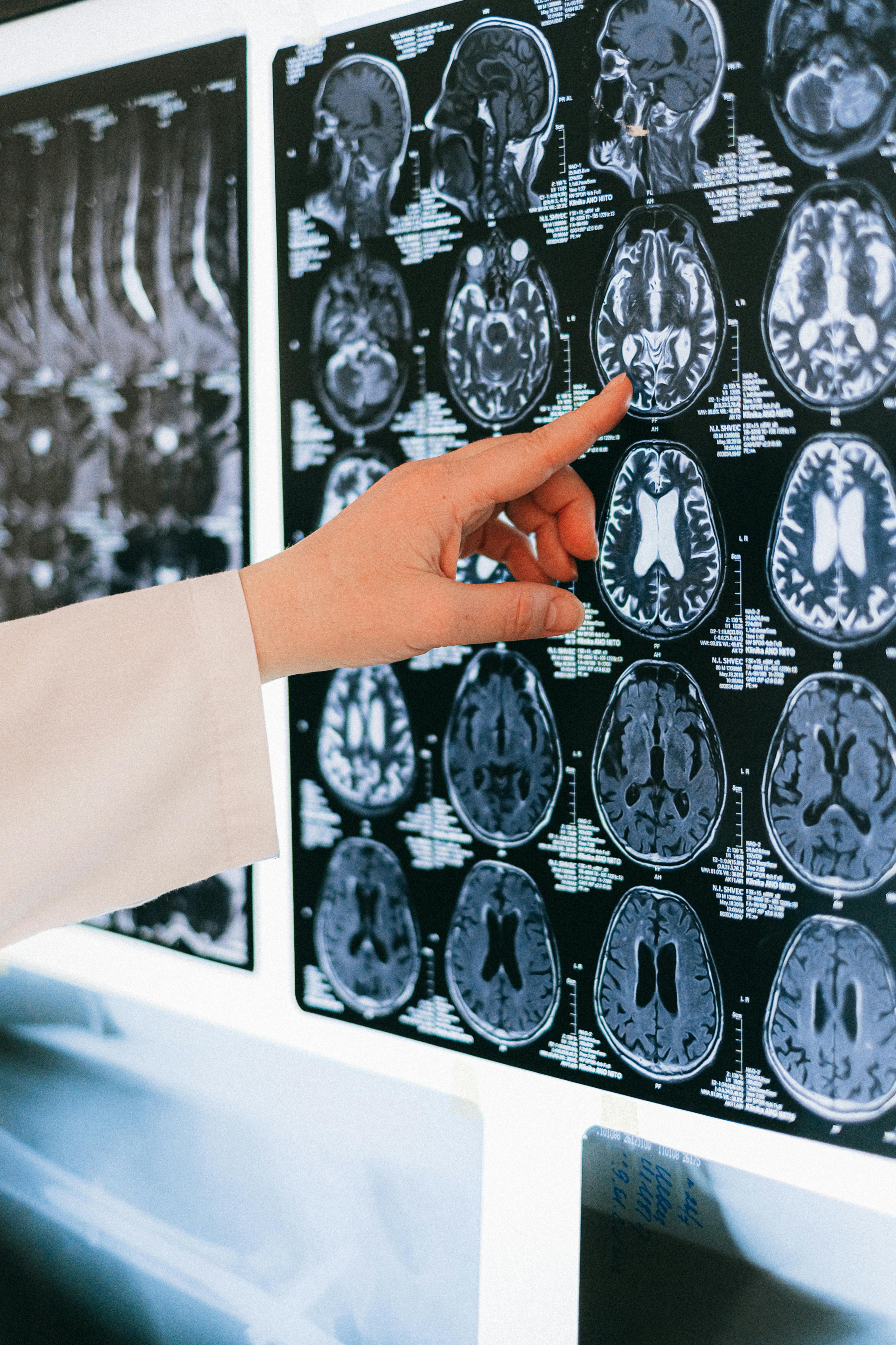
Sunday, 17 May 2026

Revolutionary brain-powered hearing lets you pick out voices in noisy crowds with ease!

Person wearing headphones in a focused setting

Have you ever been at a wedding reception, a bustling coffee shop, or a rowdy birthday party where the background noise sounds like a jumbo jet taking off inside your skull? You’re trying to listen to your friend explain their new obsession with competitive sourdough baking, but all you can hear is the clinking of forks, three other conversations about the weather, and a toddler crying in the distance. It is the age-old struggle known as the "Cocktail Party Problem," and for decades, it has been the ultimate boss fight for humans and hearing aids alike. But hold onto your hats, because science has just handed us a pair of imaginary volume knobs for our brains.

Imagine if your brain acted like a high-tech remote control. Instead of fiddling with buttons on a device, you simply look at the person you want to hear, and—presto!—their voice becomes crystal clear while the surrounding chaos fades into a soft, manageable hum. This isn’t science fiction or a scene from a superhero movie; it is the result of some seriously clever folks figuring out how to bridge the gap between our grey matter and the digital world. We are talking about real-time, brain-controlled selective hearing that actually works while you're out and about.

The secret sauce behind this wizardry involves a mix of mind-reading sensors and super-smart artificial intelligence. Traditional hearing aids have always been a bit blunt; they usually just turn the volume up on everything, which often makes the noise problem even worse. This new approach, however, uses sensors to eavesdrop on your brainwaves. When you focus on a specific speaker, your brain actually starts to "sync" its activity with the rhythm and patterns of that person's voice. It’s like your neurons are dancing to the same beat as the person talking to you. The system detects this neural choreography and says, "Aha! That’s the voice we want!"

Once the system identifies who you’re trying to listen to, the AI kicks into overdrive. It acts like a digital bouncer, grabbing that specific voice and giving it a VIP pass to your eardrums while politely but firmly showing the background noise the exit. The coolest part? This all happens in the blink of an eye. In the past, this kind of tech was sluggish, making the audio feel like a badly dubbed foreign film where the lips don’t match the sounds. Now, the lag is so minimal that it feels completely natural, like you’ve suddenly developed a superpower you never knew you had.

This breakthrough is a massive deal for anyone who has ever felt isolated in a crowd because they couldn't follow the thread of a conversation. It turns a stressful social situation into a walk in the park. But the fun doesn't stop at just helping those with hearing loss. Imagine being a student in a noisy lecture hall or a busy office worker trying to focus on a single colleague in an open-plan nightmare. This technology could eventually become a standard feature in the earbuds we wear every day, turning us all into social ninjas who never miss a punchline or a secret.

Of course, we aren't quite at the stage where we can buy "Mind-Ear 3000" at the local electronics store just yet. The current setups often involve wearing a cap or sensors that make you look a bit like an extra from a 1980s space opera. However, researchers are working hard to shrink everything down. The goal is to hide these brain-reading sensors inside sleek, everyday earphones or even tiny implants that sit discreetly behind the ear. Before you know it, you’ll be able to tune out your annoying neighbor's leaf blower just by thinking about it.

What makes this leap so exciting is how it treats the human brain as an active participant in the technology. We aren't just passive listeners anymore; we are the conductors of our own personal orchestra of sound. By harnessing the natural way our brains focus, scientists are creating tools that feel like an extension of our own bodies. It’s a beautiful marriage of biology and bits, proving that the future of hearing isn’t just about making things louder—it’s about making things smarter.

So, the next time you find yourself in a crowded room, nodding along to a story you can’t actually hear, take heart. The days of the "smile and nod" strategy are numbered. Soon, your brain will be the ultimate DJ, mixing the sounds of your life in real-time, ensuring that the voices that matter most are always front and center. It’s a brave new world of sound, and it’s going to be music to everyone’s ears.

Friday, 15 May 2026

Discovering Her True Calling and Community: An Inspiring Neuroscience Journey at George Mason!

Imagine walking onto a university campus and feeling like you’ve just stepped into the headquarters of a secret society of thinkers, dreamers, and doers. That is exactly what happened when one bright-eyed student first set foot at George Mason University. She wasn't just looking for a degree or a fancy piece of paper to hang on her wall; she was on a quest to unlock the most complex machinery in the known universe: the human brain. For her, neuroscience wasn't just a major choice made on a whim; it was a calling that echoed through every nerve ending and synapse in her body.

When she arrived at the green and gold grounds of George Mason, the air felt different. It was charged with the kind of electric energy you only get when thousands of curious minds are all trying to solve the world's puzzles at once. While some people might find the idea of studying neural pathways and chemical transmitters a bit daunting, she saw it as the ultimate adventure. To her, every lecture was a map and every lab session was a journey into the deep, dark, and utterly fascinating woods of the human consciousness. She quickly realized that she wasn't just a face in the crowd; she was a Patriot in the making, ready to dive headfirst into the "gray matter" of life.

The magic truly began when she found her "neuro-fam." We all know that university can sometimes feel like a giant ocean where you’re just trying to keep your head above water, but she found a lifeboat filled with fellow brain-enthusiasts. This community wasn't just about sharing notes or cramming for midterms over lukewarm coffee at two in the morning—though there was plenty of that, too! It was about finding people who got just as excited as she did about the way a single neuron fires or how a specific protein can change the way we remember our favorite childhood snacks. This sense of belonging turned the rigorous academic path into a collaborative celebration of discovery.

In the lab, things got even more exciting. Picture a space where high-tech gadgets and glowing screens are the norm, and where "playing with your food" is replaced by "analyzing biological data." She spent countless hours peering through microscopes and crunching numbers, fueled by a passion that most people reserve for rock concerts or sports championships. There is a certain kind of thrill that comes with seeing a theory from a textbook come to life right before your eyes. She wasn't just learning about science; she was doing it, contributing her own tiny piece to the massive jigsaw puzzle of how we perceive the world around us.

Scientific Research Laboratory

But it wasn't all just test tubes and research papers. Life at George Mason provided a vibrant backdrop for her to grow as a person, not just a scientist. Between the bustling energy of the Johnson Center and the quiet, leaf-strewn paths of the Fairfax campus, she discovered that being a student is about finding balance. She learned that a breakthrough in the lab is much sweeter when you have a group of friends to celebrate with afterward, perhaps over some local treats or a quick trip into the heart of the city. These moments of connection were the "glue" that held her academic journey together, proving that even the most brilliant brain needs a heart full of community to truly thrive.

Mentorship played a massive role in her transformation as well. Imagine having a guide who has already climbed the mountain you’re currently scaling, someone who can point out the slippery rocks and show you the best views. The faculty members weren't just distant figures behind a podium; they were mentors who saw her potential and pushed her to reach further than she thought possible. They encouraged her to ask the "weird" questions, the ones that don't have easy answers, because that is where the real growth happens. Through their guidance, she transitioned from a student who was absorbing knowledge to a researcher who was generating it.

As she nears the next chapter of her story, the future looks as bright as a firing synapse. The skills she gathered—the critical thinking, the technical prowess, and the ability to work within a team—are like a Swiss Army knife for the professional world. Whether she ends up in a high-stakes research facility, a medical setting, or an innovative tech hub, she carries with her the spirit of George Mason. She found more than just a career path; she found a version of herself that is confident, connected, and ready to take on the mysteries of the mind. It just goes to show that when you follow your curiosity and find the right tribe, the possibilities are truly mind-blowing!

Ultimately, her journey serves as a colorful reminder that university is what you make of it. It’s about more than just the grades; it’s about the "aha!" moments, the lifelong friendships, and the realization that you are part of something much bigger than yourself. She walked onto campus looking for a major, but she walked away with a mission. And in the world of neuroscience, where there is still so much left to discover, having a mission-driven mind like hers is exactly what we need to light up the future. From the classroom to the laboratory, her story is a sparkling example of what happens when passion meets opportunity in the perfect environment.

Wednesday, 13 May 2026

Amazing Discovery Reveals How Layered Brain Circuits Control Human Epileptic Activity!

Imagine your brain is a massive, multi-story nightclub that never closes its doors. It is a place where the music is always playing, and the dance floor is packed with billions of tiny guests known as neurons. For most of the night, the vibe is perfect—the rhythm is steady, and everyone is moving in sync. However, in some brains, there is a bit of a glitch in the sound system. These little electrical hiccups, known to the science crowd as interictal discharges, are like the speakers popping and crackling between songs. They are not full-blown power outages, but they are definitely strange vibes that happen when things should be chill. For a long time, the mystery was: which floor of the nightclub is causing all the noise?

It turns out that the brain is not just one big, open room. It is actually a very organized six-story skyscraper, and each floor—or "lamina"—has its own specialized team of workers and a very specific job to do. Recent deep-dives into this biological architecture have revealed that these tiny electrical sparks do not just happen randomly throughout the building. They have a favorite hangout spot. By eavesdropping on the cellular chatter, researchers discovered that the middle and upper floors are often where the drama begins. It is as if the VIP lounge on the second and third floors starts a heated argument that eventually leaks down to the lobby and disrupts the whole building.

Stylized glowing neural network representing brain activity

To understand how this works, we have to look at the "microcircuits." These are tiny groups of neurons that act like tight-knit gossip circles. In a brain that is functioning smoothly, these circles keep secrets well and keep the volume at a reasonable level. They have a system of checks and balances. But in a brain prone to these little electrical bursts, the gossip spreads way too fast. One neuron tells another, "Hey, did you hear?" and before you know it, a whole group of cells is shouting the exact same thing at the exact same time. This synchronized shouting is what creates those sharp electrical spikes that show up on medical monitors.

The really fascinating part is that the researchers found specific "peacekeeper" cells that try to hush everyone up. These cells are like the world’s most exhausted security guards. They see the shouting match starting and try to jump in to calm things down, but sometimes the "cheerleader" cells—the ones pushing for more noise—are just too loud and too fast. This tug-of-war between the shushers and the shouters happens across the different floors of the brain, creating a complex map of electrical activity that changes depending on which floor you are standing on.

How did we find this out? It was not by just looking at the outside of the building. Scientists used incredibly tiny, high-tech probes—think of them as microscopic microphones—to record the individual conversations of neurons within actual human brain tissue. This is essentially the neuroscience version of a high-stakes reality show, but with much more electricity and significantly more importance for human health. By tracking which cells fired and exactly when they did it, the team could watch the spark travel from one layer to another, finally identifying the "who-said-what" of brain glitches.

Why does all of this matter to those of us who aren't wearing lab coats? Well, if we know exactly which floor the noise is coming from and which specific group of neurons is failing to keep the peace, we can start designing much better ways to fix the problem. Instead of just shutting down the whole nightclub with heavy-duty medications that make everyone sleepy and sluggish, we might one day be able to send a specific "shusher" directly to the third floor. This kind of pinpoint accuracy is the ultimate goal for fixing brain-related hiccups without bothering the rest of the party.

This discovery that our brain layers are so specialized in handling these discharges is a massive leap forward. It shows that our grey matter is even more organized and "layered" than we previously imagined. Each floor is like a different instrument in a grand orchestra, and when one goes out of tune, the whole symphony feels the tension. By learning how to tune just those specific strings, we are getting closer to a world where the music stays smooth and the "pops" and "crackles" become a thing of the past. It is a wild, microscopic world in there, and we are finally getting the VIP tour of the circuits that make us who we are.

Sunday, 10 May 2026

Unlock the Amazing Secrets of the Human Brain with This Thrilling Neuroscience Discovery!

Neuroscience Article A colorful brain model representing creativity and sound

Ever wondered why your toes start tapping the second a funky bassline kicks in? It’s not just a reflex; it’s a full-blown rave happening inside your skull! Your brain is the ultimate music snob, but in the best way possible. It doesn’t just hear sound; it dissects it, dances with it, and weaves it into the very fabric of your memories. When you press play on your favorite playlist, you’re essentially handing your neurons a pair of glow sticks and telling them to go wild. This magical connection between melodies and grey matter is a scientific rollercoaster that researchers are finally starting to map out in vivid detail.

Think of your ears as the high-tech security guards at the entrance of a VIP club. Sound waves come knocking, and these guards translate those vibrations into electrical signals—the secret language of the brain. These signals zoom through the auditory nerve like a fleet of tiny, caffeinated messengers, heading straight for the auditory cortex. This part of your brain is the head chef, taking raw ingredients like pitch, rhythm, and timbre, and whipping them up into a delicious auditory souffle. It’s a process so fast and complex that even the world’s fastest supercomputer would get a bit of "buffer face" trying to keep up with a fast-paced jazz solo.

But wait, there’s more! Music doesn’t just stay in one corner of your mind. It spreads like glitter at a craft fair. While the auditory cortex is busy with the notes, other parts of your brain are checking the rhythm. Your motor cortex—the part that usually handles things like walking or holding a coffee cup—starts firing off signals as if it wants to join a dance troupe. This is why it’s almost impossible to stay perfectly still when a heavy beat drops. Your brain is literally preparing your muscles to move in sync with the sound, turning your entire body into a giant antenna for rhythm. It’s a biological urge to groove that dates back to our earliest ancestors drumming on hollow logs.

Why does music feel so good? That’s thanks to our old friend dopamine, the brain’s personal "victory candy." When you hear a song you love, or better yet, when a song builds up to a massive crescendo and finally drops, your brain releases a flood of this feel-good chemical. It’s the same reward system that fires off when you eat a delicious slice of pizza or win a game of your favorite video game. Scientists have found that the brain actually anticipates these musical peaks, creating a sense of delicious tension and release that keeps us hooked. It’s a natural, healthy high that requires nothing more than a decent pair of headphones and a dream.

Have you ever heard a song from ten years ago and suddenly remembered exactly what you were wearing, who you were with, and even what the air smelled like? That’s because music is the ultimate time machine. The hippocampus, which acts as the brain’s librarian, stores musical memories in a very special, high-security vault. Because music is tied so deeply to our emotions, these memories are etched in high definition. This is why music therapy is such a powerhouse tool for people dealing with memory loss; even when other memories begin to fade, the "soundtrack" of their lives often remains crystal clear, ready to be played back with a single familiar chord.

Beyond just individual vibes, music is a social glue that has been sticking humans together for millennia. When we sing in a choir, jam in a band, or dance at a massive concert, our brain waves actually start to synchronize with the people around us. It’s a phenomenon called neural coupling. We begin to feel what others feel, creating a shared emotional experience that’s hard to find anywhere else. It’s like our brains are all logging into the same Wi-Fi network, sharing data and vibes in real-time. This collective harmony is likely why music has been a part of every single human culture since we first discovered we could make noise with our throats and hands.

Because music engages so many different parts of the brain at once—the areas for emotion, movement, language, and memory—it’s being used as a secret weapon in modern medicine. For someone who has trouble speaking after a stroke, singing can sometimes bypass the damaged language centers and allow them to communicate through melody. For those with movement disorders, a steady, driving beat can act as an external metronome, helping them find their stride again. It turns out that a song isn't just a song; it's a multi-tool for the human mind, capable of repairing, soothing, and energizing us in ways we are only just beginning to fully understand.

So, the next time you find yourself getting lost in a symphony or air-drumming in your car at a red light, remember that you’re participating in one of the most complex biological performances on Earth. Your brain is a masterpiece of engineering, tuned specifically to the frequency of joy. Keep those headphones handy and your playlists diverse, because your neurons are always ready for an encore. Whether it’s heavy metal, smooth jazz, or catchy pop, your brain is the most dedicated fan in the front row, cheering for every single note and every single beat. Science proves it: we are literally built to rock and roll!

Friday, 8 May 2026

Incredible Breakthrough Reveals How Sugar Powers the Birth of New Brain Cells!

Imagine your brain is a giant, bustling metropolis, filled with millions of tiny electrical wires zipping messages from your toes to your nose and everywhere in between. To keep these messages moving at lightning speed, the wires need some serious insulation. Enter the unsung heroes of the neural neighborhood: the Oligodendrocyte Progenitor Cells, or as their friends call them, the OPCs. These little guys are like the enthusiastic junior apprentices of the brain’s construction crew. Their primary job is to grow up, settle down, and wrap those neural wires in a cozy, fatty blanket called myelin. Without this insulation, our internal internet would be about as fast as dial-up in a thunderstorm. But here is the kicker: these apprentices need to know exactly when and where to start multiplying to keep the city running smoothly.

Scientists have recently discovered that these tiny builders are actually massive foodies. It turns out that their favorite snack—glucose—is much more than just a source of energy. For an OPC, a surge of sugar is like receiving a high-priority work order from the mayor. When glucose levels are just right, these cells get the signal to start a "progenitor party," dividing and multiplying like crazy to ensure there are enough workers on the site. This isn't just a random occurrence; it’s a finely tuned process that changes depending on where the cells are located in the brain and what time of day it is. It’s as if the brain has its own internal GPS and clock telling the builders, "Hey, we need more insulation in the prefrontal cortex at 2:00 PM, so grab a snack and get to work!"

Colorful representation of a neural network

How does a simple sugar molecule tell a cell to start making copies of itself? This is where a very special "chef" called ACLY comes into play. ACLY, or ATP citrate lyase for those who like the long version, is an enzyme that acts as a master translator. It takes the energy coming in from the glucose and converts it into a specific chemical currency. This currency is then used to decorate the cell's DNA in a process known as histone acetylation. Think of histone acetylation as the cell’s way of using bright neon highlighters on its own instruction manual. When the ACLY chef provides enough "ink," the cell highlights the chapters titled "How to Multiply" and "Building More Builders."

This "neon highlighting" is a game-changer because it physically changes how the DNA is packed inside the cell. When the glucose is flowing and ACLY is busy at work, the DNA relaxes a bit, making it easier for the cell to read the blueprints for growth. This isn't just a one-size-fits-all situation, though. The study revealed that this process is incredibly sensitive to the environment. In some parts of the brain, the cells are much more responsive to these sugar-coated instructions than in others. This spatial modulation ensures that the brain isn't just throwing insulation at random spots but is targeting the areas that actually need the most maintenance.

The temporal aspect—the "when"—is just as fascinating. Our bodies follow natural rhythms, and it seems our brain's construction crew follows a strict schedule too. The availability of glucose and the activity of the ACLY enzyme fluctuate, creating windows of opportunity for the OPCs to expand their workforce. By syncing their growth with the body's metabolic clock, these cells ensure they are operating at peak efficiency. It’s like a smart factory that only runs the assembly line when power is cheapest and the most materials are available. This level of organization is what keeps our brains adaptable and capable of repairing themselves after a long day of thinking, learning, and scrolling through memes.

Why does all of this matter to us? Well, understanding how these "sugar signals" work opens up a world of possibilities for brain health. Sometimes, the construction crew goes on strike or runs out of supplies, which happens in conditions like multiple sclerosis or as we get older. In these cases, the neural wires lose their insulation, and the messages start to lag. By figuring out exactly how glucose and ACLY tell OPCs to multiply, scientists might be able to jumpstart the repair process. Imagine being able to give the brain’s builders exactly the "menu" they need to get back to work, wrapping those wires and restoring the brain’s high-speed connection.

In the end, it’s a beautiful reminder that everything in our bodies is interconnected. A bit of sugar isn't just fuel for a run or a sweet treat for your tongue; it’s a sophisticated messenger that helps shape the very structure of your mind. The next time you feel a "sugar rush," just imagine those tiny OPC apprentices in your brain, highlighter pens in hand, getting ready to build a better, faster, and more insulated you. It’s a sweet deal for everyone involved!

Thursday, 7 May 2026

Unlocking the Neuroscience of Reward: How a Newly Discovered Neural Network Drives Eating Behaviors

The human relationship with food is notoriously complex. We may feel completely satiated after a large meal, yet still find ourselves reaching for a sugary dessert. At other times, our bodies might intensely crave salty snacks or protein-dense meals. For decades, neuroscientists have sought to understand the exact mechanisms that govern these shifting desires.

Now, groundbreaking research from the University of Delaware provides a crucial missing link. By studying the microscopic brains of fruit flies, researchers have identified a specific neural network responsible for the earliest stages of value computation—the process by which the brain decides whether a specific taste is rewarding in any given moment.

Published in the prestigious journal Current Biology, this research, led by Dr. Lisha Shao, an assistant professor in the Department of Biological Sciences at the University of Delaware’s College of Arts and Sciences, offers profound insights into the mammalian reward system. By mapping these fundamental pathways, scientists are gaining a clearer understanding of how healthy eating habits are formed, and conversely, how neurological misfires can lead to severe conditions such as binge-eating, anorexia, and addiction.

A 3D scientific visualization of a glowing neural network demonstrating brain synapses firing.

The Mechanics of Value Computation

Historically, scientists have possessed a strong understanding of the mechanics of taste. When we consume food, sensory neurons in our taste buds detect basic flavor profiles—sweet, salty, bitter, sour, and umami—and transmit these signals to the brain. However, sensory detection is only the first step.

The more complex neurological hurdle has been understanding how the brain interprets those signals. A donut is inherently sweet, but its reward value fluctuates drastically depending on the consumer's current state.

According to Dr. Shao’s research, the brain does not assign a static value to food. Instead, it engages in dynamic value computation, which integrates three primary factors:

  • Internal Physiological State: Is the organism hungry, satiated, or lacking specific nutrients?
  • Environmental Context: Is the food safe to eat, and what are the immediate surroundings?
  • Past Experience: Has this food caused illness or provided sustained energy in the past?

"Our goal is to understand how the brain assigns value—why sometimes eating something is rewarding and other times it’s not," Shao notes. "If the brain assigns the wrong value to something—too much or too little—behavior goes wrong."

Fox Neurons: The Gatekeepers of Reward

To isolate the exact moment value computation begins, Dr. Shao’s team turned to Drosophila melanogaster, the common fruit fly. While a fruit fly's brain is no larger than a pinpoint, it relies on many of the same foundational chemical messengers and structural building blocks as the human brain. Remarkably, humans and fruit flies share approximately 60% of their disease-related genes, making these insects an invaluable model organism for mapping complex neurological circuits.

During their experiments, the researchers pinpointed a specific pair of cells dubbed Fox neurons—named for their distinct resemblance to the pointed ears of a fox.

The researchers discovered that these Fox neurons act as the earliest known site in the fly's brain where the value computation for taste is processed. When the team artificially activated these neurons, the behavioral change in the flies was dramatic: they consumed significantly larger quantities of food, even if they had just been fed to satiation.

Furthermore, the research demonstrated that flies adjust their dietary preferences based on acute biological needs, a process entirely mediated by these neural circuits. For instance:

  • Reproductive Females: Exhibited a strong preference for protein-rich foods, which are essential for egg production.
  • Males and Non-Reproductive Females: Maintained a balanced intake of both sugars and proteins, reflecting their baseline metabolic requirements.

This targeted behavioral shift proves that the brain does not just blindly seek calories; it actively computes the nutritional value of a taste based on immediate physiological demands.

A macro photograph of a fruit fly in a laboratory setting used for genetic and neurological research.

The Evolutionary Mismatch in a Modern World

Understanding the foundational circuitry of the reward system is increasingly urgent in the modern era. From an evolutionary standpoint, the human brain evolved in environments where calorie-dense foods—particularly those high in sugar and fat—were incredibly scarce. Consequently, our neural pathways were hardwired to assign a massively high reward value to these rare resources to ensure survival.

Today, however, we face an evolutionary mismatch. The ancient neural circuits that kept our ancestors alive are now bombarded by stimuli they were never designed to process.

"Our brains evolved to process natural rewards like food and reproduction," Shao explains. "But now we’re surrounded by artificial rewards—endless short videos, processed foods—that the brain was never designed to handle."

When the brain is constantly flooded with hyper-palatable processed foods or the rapid-fire dopamine hits of social media algorithms, the value computation system can become dysregulated. The brain begins to assign disproportionately high values to harmful stimuli, laying the neurological groundwork for addiction, compulsive behaviors, and eating disorders.

Moving Beyond the "Chemical Soup"

For decades, the standard psychiatric approach to treating mood, eating, and reward-processing disorders has relied on systemic medications that alter the brain's primary chemical messengers, most notably dopamine and serotonin.

While these treatments can be life-saving, they are notoriously imprecise. Because they affect the entire brain rather than specific malfunctioning circuits, they often result in a "chemical soup" approach.

  • The Problem with Systemic Drugs: If a patient has excessively high dopamine activity driving an addiction, traditional medications lower dopamine levels globally across the brain.
  • The Consequence: This global reduction can blunt all emotional responses, causing severe side effects like lethargy, depression, and a loss of general motivation (anhedonia).

Dr. Shao’s mapping of specific circuits like the Fox neurons represents a paradigm shift toward targeted neuro-therapeutics. By understanding the exact physical pathways where decisions and value assignments are made, future medical interventions could theoretically target only the misfiring circuits, leaving the rest of the brain's delicate chemical balance undisturbed.

"If we understand how decisions are made at the circuit level, we’re one step closer to understanding why they sometimes go wrong, and how to fix them," Shao states. "You can’t fix what you don’t understand."

Ultimately, this research bridges the gap between microscopic cellular biology and complex human behavior. By decoding the tiny brain of the fruit fly, neuroscientists are moving closer to a future where eating disorders and behavioral addictions can be treated not with blunt chemical instruments, but with precise, circuit-level precision.

The Architecture of Memory: Why the Brain’s Hippocampus Begins as a 'Full Slate' Rather Than a Blank Canvas

For centuries, philosophers and early psychologists operated under the assumption that the human mind begins as a tabula rasa—a blank slate. According to this deeply entrenched theory, we are born with an empty cognitive canvas, and our experiences gradually paint the intricate neural connections that form our memories, personality, and knowledge. However, modern neuroscience is fundamentally rewriting this narrative.

A groundbreaking study published in the journal Nature Communications reveals that the brain’s primary memory center does not start empty. Instead, it begins as a tabula plena—a "full slate" characterized by a dense, hyperconnected web of neurons. It is only through the rigorous process of maturation and experience that these chaotic connections are pruned away to form the precise, efficient networks required for distinct memory recall.

3D rendering of a human brain with a glowing hippocampus

Decoding the Hippocampus and the CA3 Region

To understand how memories are built, researchers must look deep into the hippocampus, a seahorse-shaped structure nestled within the brain's temporal lobe. The hippocampus is the biological engine of memory consolidation, responsible for transforming fleeting daily experiences into long-term memory storage.

Within the hippocampus lies a highly specialized region known as the cornu ammonis 3 (CA3). The CA3 is critical for a cognitive process called pattern completion—the ability to recall an entire memory from a partial cue, such as remembering a childhood home just by catching the scent of a specific blooming flower.

The function of the CA3 relies heavily on neural plasticity, which is the brain’s ability to continuously strengthen or weaken the connections between neurons, known as synapses. When neurons fire together frequently, their synaptic connections strengthen, effectively encoding a memory. But the question that has long puzzled neuroscientists is how these CA3 networks are structured at the very beginning of life.

From Hyperconnected Chaos to Sculpted Precision

To solve this mystery, a team of researchers led by neuroscientist Peter Jonas at the Institute of Science and Technology Austria analyzed the microscopic architecture of the mouse hippocampus. By examining brain tissue at three distinct developmental stages—shortly after birth, during adolescence, and in adulthood—the researchers were able to map the lifecycle of memory networks.

The findings completely upended the tabula rasa hypothesis.

In the neonatal brain tissue, the researchers discovered that the hippocampal networks were overwhelmingly dense. Neurons within the CA3 region were hyperconnected in a seemingly random, haphazard pattern. Even more surprising was the strength of these early connections. Conventional wisdom suggested that infant brains would possess weak, underdeveloped synapses that required time to grow. Instead, the researchers found that these early synapses were incredibly robust and highly excitable.

Split view comparing dense early neural networks with sparse mature neural networks

As the brain matures from infancy into adolescence, a biological phenomenon known as synaptic pruning takes over. The brain begins to actively dismantle the excess connections. The haphazard, dense network becomes sparser, highly organized, and meticulously structured.

The developmental trajectory of memory networks can be understood through three distinct phases:

  • Neonatal Hyperconnectivity: The infant brain features a dense, highly excitable web of neurons. A single sensory input can cause a neuron to fire, leading to broad, overlapping waves of brain activity.
  • Adolescent Synaptic Pruning: As the organism interacts with its environment, the brain identifies which connections are useful and which are redundant. Unused pathways are chemically dismantled, making the network sparser but more efficient.
  • Mature Specificity: In adulthood, neurons become highly selective. They require multiple, specific inputs to fire. This selectivity allows the brain to store distinct, separate memories without them bleeding into one another.

Solving the Mystery of Infantile Amnesia

This transition from a dense network to a sparse one provides a compelling biological explanation for infantile amnesia—the universal human inability to recall memories from the first few years of life.

Because the neonatal CA3 region is so hyperconnected and excitable, different experiences trigger overlapping patterns of neural activity. If the neural activity for "eating a strawberry" looks almost identical to the neural activity for "playing with a block," the brain struggles to separate the two events. The memories are generated, but they are incredibly broad and lack specific details.

This lack of precision heavily influences behavior. In behavioral studies, when young rodents are given a mild shock in a specific corner of a cage, they learn to fear the event. However, because their neural networks lack specificity, they will freeze in fear when placed in any similar environment. The memory of the fear exists, but it is generalized. In contrast, adult rodents—whose brains have undergone synaptic pruning—will only freeze in the exact location where the shock occurred. Their refined neural networks allow for highly specific memory recall.

Therefore, we likely do not remember our infancies because those early experiences were recorded on an overly sensitive, hyperconnected system that could not file memories into distinct, retrievable categories.

Conceptual artwork of a glowing block being sculpted into a precise neural network

The Interplay of Genetics and Experience

If the brain does not build these dense early networks through experience, where do they come from? The emerging consensus is that the initial, hyperconnected architecture of the brain is driven by a genetically programmed developmental blueprint.

Before birth, our DNA dictates the rapid generation of neurons and synapses, ensuring that the organism enters the world with a "full slate," biologically prepared to absorb massive amounts of sensory data. Once born, the environment takes over as the sculptor. Postnatal experiences dictate which of those prewired connections will survive and which will be pruned away.

Dr. Hauður Freyja Ólafsdóttir, an assistant professor at the Donders Institute for Brain, Cognition and Behaviour, notes that these circuit-level discoveries align perfectly with decades of developmental psychology. While prenatal experiences and early infancy leave indelible psychological traces, they rely on a fundamentally different, less refined neural architecture than the precise memories formed later in life.

Ultimately, the brain is not a blank hard drive waiting for data to be written onto it. It is more akin to a dense block of marble. Experience does not simply add to the brain; rather, experience acts as a chisel, chipping away the excess stone to reveal the sharp, highly defined structures of our memories.

Source: https://www.livescience.com/health/neuroscience/the-brains-memory-center-doesnt-start-as-a-blank-slate-study-suggests